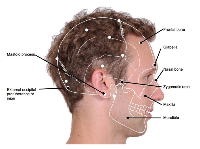
exproperti.com – The mastoid foramen is present in both the male and female skulls. It is found on the right side, while it is absent on the left side. This unusual feature is important for neurosurgery. Its multiple origins, evolutionary implications, and clinical implications are discussed. In this article, we describe the anatomy of the mastoid foramen. Here, we also discuss its neurosurgical importance.
A foramen in the skull that is located in the temporal bone
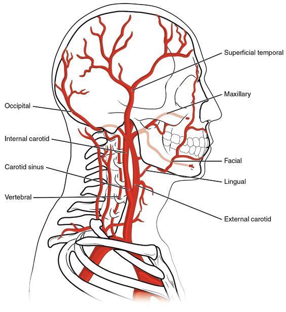
The mastoid foramen is a variably-present foramen in the skull. Its diameter and size vary, but they are always located in the temporal bone. The foramen transmits emissary veins to the sigmoid sinus and a small dural branch of the occipital artery. Anatomy of the foramen is important to medical practice.
This foramen is usually found on one side of the skull, although some patients have asymmetrical distribution. The mastoid emissary vein is usually present on both sides of the head, and the foramen is located on the left side. MRIs of the brain may be useful for diagnosing this condition. However, further research is needed to determine if this type of foramen is actually a part of the body.
The size and shape of the mastoid foramen vary in men and women. The length of the foramen varies from side to side. It is most commonly narrow on one side, and wide on the other. It is located below the labyrinth, and the distance from the sigmoid sinus to the outermost part of the skull is the same for both. The circumference of the foramen is the same in men and women.
Anatomical markers are important in determining the genetic relationships of populations
The mastoid foramen is a narrow, curved, and irregular foramen. The foramen is also a vital anatomic landmark in determining the genetic relationships of populations. Its location on the skull is highly significant for forensics, anthropology, and other fields. When there is a small foramen, it can indicate the presence of a bone-like structure.
The anatomy of the mastoid foramen has significant importance for forensic and plastic surgery. It is an anatomical landmark used to differentiate different species. Various anatomical landmarks have been identified. These include the occipital emissary vein, the ibis, and the emissary vein. In many cases, the foramens are not symmetrical.
The foramen is a source of pain, swelling and abscess
The mastoid foramen transmits a blood vessel and nerve. It is anatomically similar to the occipital foramen. The foramen in the temporal region is often enlarged. The foramen is a source of pain, swelling, and abscess. In these cases, the occipital artery and emissary vein are the most common causes of traumatic headaches.
A mastoid segment gives off a sensitivity branch to the auricular area. It starts at the facial canal and extends five millimeters anteromedially. It is bounded by the middle and posterior fossa dura plates. A lateral foramen lodges the lesser superficial petrosal nerve. These vessels give off anatomical variations. These foramina are positioned on either side of the mastoid horn.
The mastoid foramen is an opening at the posterior border of the temporal bone
Anatomically, the mastoid foramen is a hole at the temporal bone’s posterior edge. It connects the emissary vein to the dura mater, and is a critical anatomical landmark in forensic science. It can be easily visualized in anatomical views. Its size and position vary. The mastoid foramen is usually intracranial, although sometimes it is located at the suture between the occipital and temporal bone.
Anatomical differences between human and nonhuman animals can be explained by the shape of the foramen. In the human skull, the mastoid emissary vein is located in the temporal bone, while the mastoid foramen is located at the temporal bone. Scientists have hypothesized that the emissary vein is the main source of blood flow in the skull. The brain is a crucial part of the body.
The tympanic segment starts from the first genu and turns backward. It lies in a thin-walled bony canal between the lateral SSC and the incus. In male skulls, the tympanic segment forms an angle of 95-125 degrees with the mastoid foramen. The tympanic vein is the main source of blood flow in the human skull.

