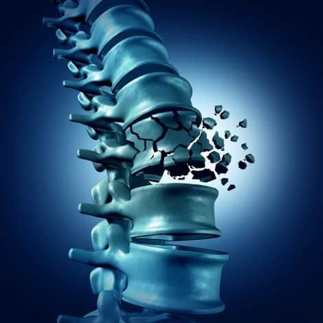
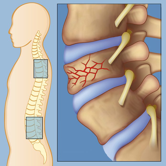
Exproperti.com – Anterior wedge fracture occurs when the vertebral body is flexed forward from its normal position. This causes a disruption in the posterior axis, resulting in axial height loss. The posterior elements of the spine then subluxate inferiorly, distracting the zygapophysial joint. A periosteal irritation and pseudarthrosis may result.
In cadaveric studies, rigid materials (PMMA) are used to obtain wedge fractures

There is no evidence that forward flexion loading is directly responsible for an anterior wedge fracture. Furthermore, there is no evidence to support the notion that morphometry and material properties of the disc play a role in the formation of a wedge fracture. In cadaveric studies, stiff material (PMMA) is used to elicit a wedge fracture. Hence, the risk of an anterior wedge fracture remains low.
This fracture involves the two posterior elements, the inferior and superior articular processes. During flexion of the vertebra above the fractured one, the inferior articular process will be drawn cephalad. Because the inferior articular process hangs from the superior articular process, it can cause instability. In addition, it is not common to experience a second-degree posterior wedge fracture in a person who has been involved in a recent fall.
The morphological changes are similar to compression fractures

Anterior wedge fracture is difficult to differentiate from physiological wedging. In an adult, the anterior vertebral body height is less than 20% of its normal value. However, anterior wedge fracture at the L5 vertebra does not have this characteristic. It is more likely to be a compression fracture in which the endplates are preserved. The morphological changes are similar to a compression fracture. Despite the complexity of diagnosis, it has important prognostic implications.
The severity of the injury is often not determined in the first visit. However, if the patient is neurologically intact and does not require surgery, treatment options may include nonoperative treatments. Nonoperative treatments include bed rest and activity limitations for a few weeks, physical therapy, and pain medications. Osteoporosis fracture prevention may include the use of bisophosphonates, calcium, and exercise. If the patient has an anterior wedge fracture, a surgical treatment may be indicated.
Physical activity is very important for spinal fracture prevention

While osteoporosis is the most common cause of vertebral fractures, there is no cure for osteoporosis, which is why physical activity is so important for the prevention of a vertebral compression fracture. Despite the benefits of physical activity, people with osteoporosis often fear exercising, because they believe it will worsen their condition. Fortunately, there are many exercise programs that can help individuals with painful vertebral fractures stay active.
Although the incidence of this fracture varies by gender, it is the most common type of vertebral fracture. Age-adjusted rates were higher in women than men. More than 90% of cases involved trauma of some sort, with a higher incidence in women. In general, the incidence of fractures following moderate trauma occurred in women, while men had a lower incidence of this type of fracture. But, the study did not specify whether the fracture was caused by trauma or age-related.
Patients with a diagnosis of a chest compression fracture have a higher risk of death

After a diagnosis of an anterior wedge fracture, the patient may experience exaggerated lordosis and kyphosis. These injuries can also decrease the patient’s exercise tolerance and abdominal space. They may also suffer from sleep disorders, decreased self-esteem, and an impaired ability to perform daily activities. Patients with a diagnosis of a thoracic compression fracture are at a higher risk for death and reduced quality of life.
Biomechanics explains how vertebrae are affected by the loss of height in the anterior limb. Hence, the posterior elements must tilt or sublux according to the loss of height in the anterior pillar. While this theory cannot explain the biomechanical cause of pain in all cases, it provides an explanation for some cases. This model can be validated through clinical trials. Its limitations, however, are not as well-defined as biomechanics.


